










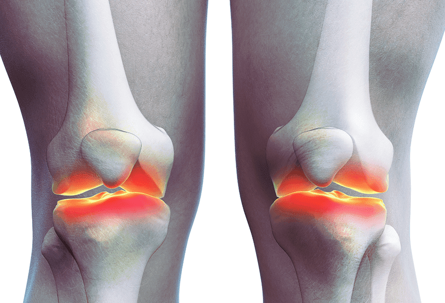








In severe cases, the condition can be identified from an X-ray alone. As the cartilage wears away, the space between the bones narrows, so the X-ray shows a narrowed joint space; as the disease progresses, bone spurs form along the edges of the knee. Loose bodies (bone fragments) may float inside the knee, and bone cysts (holes inside the bone) may form. In less severe cases, detailed tests such as MRI can show exactly how much cartilage has worn away.
 Stage 1
 Stage 2
 Stage 3
 Stage 4
 [Medication]
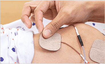 [Injection Therapy]
 [Physical Therapy]
 [Manual Therapy]
 [Shockwave Therapy (ESWT)]
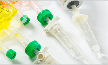 [Prolotherapy]
 [Knee Arthroscopy]
 [High Tibial Osteotomy]
 [Stem Cell Cartilage Regeneration]
 [Total Knee Replacement]


