





Dangol Hospital

Dangol Hospital
HOME 한국어
  • Conditions
  • Shoulder
  • Calcific Tendinitis
  • Conditions

    Learn about the symptoms, causes, self-check and treatment of calcific tendinitis.

    Dangol Hospital logo

    M E A N I N G

    What Is Calcific Tendinitis?

    A condition in which calcium deposits in a shoulder tendon harden like stone,
    causing severe pain.

    The exact cause is unknown, but calcium deposits are thought to form
    because of overuse of the shoulder, degenerative changes in the tendon,
    reduced blood supply and the process of calcium removal.
    The deposits vary widely in size, from 1 to 3 mm in diameter, and one or several
    may form at once. The pain is often so severe that patients cannot sleep at night,
    and many cases are discovered after a visit to the emergency room.

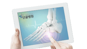
    Shoulder X-ray showing a calcium deposit

    SYMPTOMSymptoms of Calcific Tendinitis

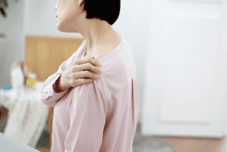
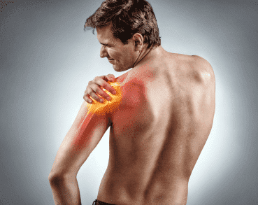
    Woman holding her painful shoulder 01

    Intense pain when raising or rotating the arm

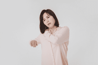
    Woman with shoulder pain 02

    Pain that varies widely, from mild discomfort to sudden severe pain

    Woman brushing her hair 03

    Difficulty with everyday movements that use the shoulder, such as dressing or brushing your hair

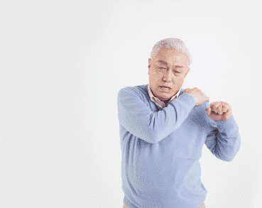
    Older man holding his shoulder 04

    Shoulder pain without any particular injury

    Woman unable to sleep 05

    Pain so severe that it prevents a good night's sleep

    CAUSECauses of Calcific Tendinitis

    Degenerative changes in the tendons due to aging

    Poor blood circulation caused by overuse of the shoulder

    Tendon cells change into cartilage cells and calcium is deposited

    SELF-DIAGNOSISCalcific Tendinitis Self-Check

    I have difficulty rotating my arm.

    The pain gets worse at night and disturbs my sleep.

    The pain starts at the front of my shoulder and spreads to my neck or arm.

    It hurts when I press below the acromion (the bony tip of the shoulder).

    Over time, the pain gets worse and the area swells.

    ※ If even one of these applies to you, we recommend consulting a specialist for an accurate diagnosis and appropriate treatment.

    DIAGNOSIS

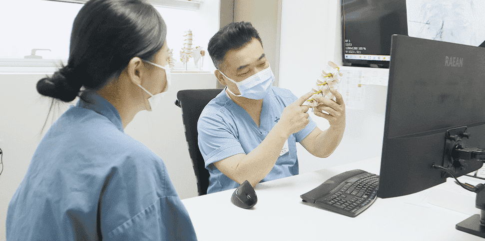
    Doctor explaining a diagnosis with a joint model

    DIAGNOSISDiagnosis at Dangol Hospital

    In most cases, the pain is so severe that patients cannot sleep,
    and their quality of life drops sharply.
    It can be treated with pain-relief treatment and rehabilitation exercise,
    and it can be diagnosed with a simple X-ray, so if you notice symptoms,
    it is best to see a doctor.
    TREATMENT

    Treating
    Calcific Tendinitis

    It is important to relieve the initial pain with anti-inflammatory painkillers, muscle relaxants or injections, so that the patient can carry out exercise therapy on their own. Most cases improve with non-surgical treatment, but in rare cases where symptoms last too long, surgery may be performed.

    CORE OF TREATMENTKey to Treating Calcific Tendinitis

    Pain can usually be relieved rapidly with a treatment of about 10 minutes,
    and rehabilitation to relax the muscles around the joint and strengthen them is very effective.

    Step 1

    In most cases, 2–3 weeks of non-surgical treatment such as medication and physical therapy brings improvement. If not, treatment moves on to injection therapy and manual therapy.

    Step 2

    Surgery is the last resort. Before considering surgery, we provide customized procedures based on each patient's condition and circumstances.

    Step 3

    Surgery may be considered for patients whose pain persists despite sufficient treatment or who need emergency care. The surgical approach and expected outcome are decided in consultation with a specialist.

    Step by StepCalcific Tendinitis Treatment at a Glance

    Step 1: Conservative Treatment

    In most cases, 2–3 weeks of non-surgical treatment such as medication, physical therapy and pain injections brings improvement.
    If not, treatment moves on to injection therapy and manual therapy.

    Patient receiving medication [Medication]

    Anti-inflammatory painkillers and muscle relaxants relieve pain and make it easier to continue physical therapy.

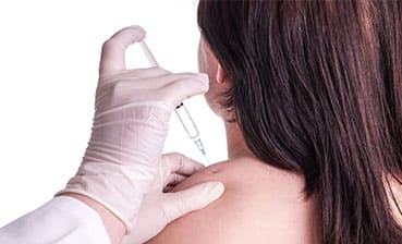
    Injection into the shoulder [Injection Therapy]

    Steroid medication or trigger point injections (TPI) are injected into the areas of muscle causing pain to relax the contracted muscles. This treatment can be tried in the early stages.

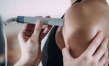
    Physical therapy on the shoulder [Physical Therapy]

    Uses heat, air, light, electricity, ultrasound and other methods to restore function around the painful area and support rehabilitation.

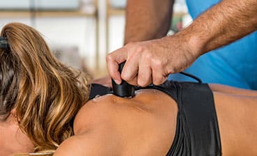
    Therapist performing manual therapy on the shoulder [Manual Therapy]

    A form of physical therapy performed by hand by a specially trained manual therapist, based on knowledge of human anatomy and physiology.

    Shockwave therapy on the shoulder [Shockwave Therapy (ESWT)]

    Extracorporeal shockwave therapy treats musculoskeletal pain from outside the body using a device that generates shockwaves.

    Step 2: Non-Surgical Procedures

    Surgery is the last resort. Before considering surgery,
    we provide customized procedures based on each patient's condition and circumstances.

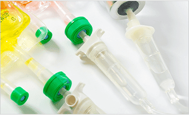
    Prolotherapy injection solutions [Prolotherapy]

    A high-concentration dextrose solution, harmless to the body, is injected directly into weakened ligaments or tendons.

    Learn More
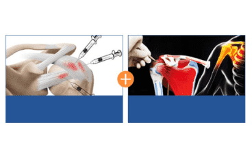
    FIMS therapy combines prolotherapy and functional intramuscular stimulation
    What Is FIMS Therapy?
    Prolotherapy
    (Proliferation Therapy)
    FIMS (Functional Intramuscular
    Stimulation)
    A non-surgical treatment combining both!
    [FIMS Therapy]

    A non-surgical treatment that combines prolotherapy (proliferation therapy) with FIMS (Functional Intramuscular Stimulation).

    Learn More

    Step 3: Surgical Treatment

    Surgery may be considered for patients whose pain persists despite sufficient treatment or who need emergency care.
    The surgical approach and expected outcome are decided in consultation with a specialist.

    Illustration of shoulder arthroscopy [Shoulder Arthroscopy]

    Through minimal incisions of less than 1 cm, an arthroscopic camera and surgical instruments are inserted into the joint to diagnose and treat the condition.

    Learn More
    Book Online
    Naver Booking
    KakaoTalk
    Chat
    Doctors & Hours
    Location
    Dangol
    SNS
    TOP